
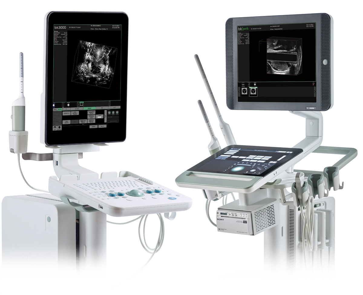
THE INFORMATION YOU NEED, IN REMARKABLE DETAIL
Advanced ultrasound imaging for anorectal and pelvic floor exams
With high-definition, detailed images, advanced 3D architecture, and an enhanced, intuitive workflow, our ultrasound imaging technology helps you visualize normal and abnormal anorectal and pelvic floor anatomy—in remarkable detail.
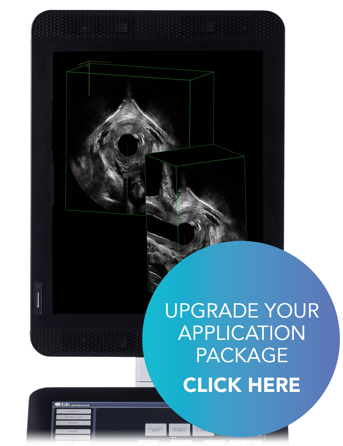
ADVANCED 3D ARCHITECTURE, WITH NEW TOOLS AND FEATURES
Easily manipulate 3D datasets
- Manipulate 3D datasets to see detailed structures from any angle you choose
- Angle and cut planes intuitively
- Compare 3D datasets by having several datasets open simultaneously
Maintain control with advanced tools and features
- Easily navigate different tools and functions
- Label 3D datasets for easy reference
- Move easily between render mode and non-render mode
Enhanced user experience
- Incorporated help functionality
- Advanced measurements and annotations
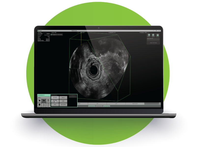
INTRODUCING THE NEW BKVIEWER!
What if there was a 3D ultrasound software that allowed you to review, manipulate, and save 3D datasets in DICOM format—all offline? Discover it for yourself today with our free trial.
DISCOVER THE BENEFITS OF BKVIEWER
3D ULTRASOUND DATA, MADE SIMPLE
-
REVIEW PLANES INTUITIVELY
Choose, manipulate, and angle planes with the intuitive controller.
-
ASSESS SLICE BY SLICE
Use the multi-slice view to step easily through a 3D cube and assess one slice at a time.
-
EASILY EXPORT DATASETS
Export 3D datasets to 3D-compatible PACS systems, and incorporate your datasets into presentations, training tools, and more.
-
DICOM-COMPLIANT
Save 3D datasets in a DICOM-compliant format and convert L3D files to DICOM.
bkViewer is not for diagnostic use.